Neuroinflammation: a silent fire
Pain medicine and rehabilitation
Neuroinflammation: a silent fire
In the first century AD, Aulus Cornelius Celsus described four signs of inflammation: redness, swelling, heat and pain. A fifth was added later, loss of function. For almost two thousand years this formula was enough to recognise inflammation by sight, without looking inside the body.
And then it turned out that in the nervous system inflammation works perfectly well having lost three of the five signs. No redness, no swelling, no heat. The blood count is calm, the ESR is normal, C-reactive protein does not rise. Only pain and loss of function remain, and both are invisible to a standard work-up. The person says that something is wrong, and the laboratory says that they are healthy.
This is exactly what neuroinflammation is: an inflammatory process inside the nervous system itself, in the spinal cord and the brain, where the work is done not by the leukocytes we are used to but by the resident cells of nervous tissue. It does not look like an abscess or like arthritis. It can be compared to smouldering wiring inside a wall, where there is neither flame nor smoke, but the light keeps flickering, and in the end goes out altogether.
The theory of neuroinflammation
Microglia make up roughly every tenth cell of the brain. They are macrophages, but not quite ordinary ones: they arrive in the brain during fetal life, from the yolk sac, and from then on live their own separate life there, almost never renewed from the bone marrow. In the biographical sense they are older than we are.
At rest, a microglial cell looks like a small body with long branching processes that continuously palpate the surrounding space. It checks whether all is well with the neighbouring neurons. During brain development it also prunes the surplus synapses, working as the gardener of neural networks. This is an important detail: microglia are not only the immune guard, they take part in construction.
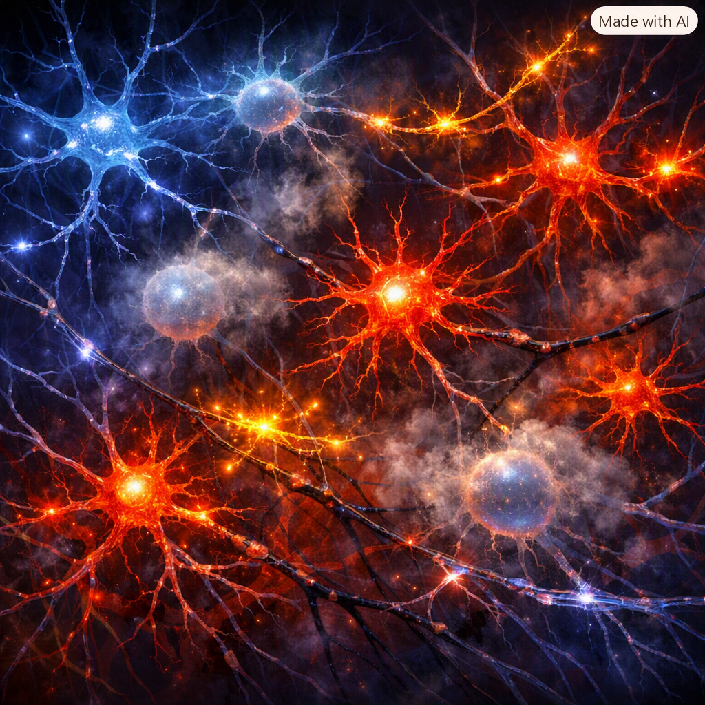
Under alarm, microglia change physically. The cell body enlarges, the processes shorten, and the cell switches into a mode of releasing pro-inflammatory substances: interleukin-1 beta, tumour necrosis factor alpha, interleukin-6, reactive oxygen species. Astrocytes join in alongside. All of this together is called glial activation.
For a short episode this is an ideal protective mechanism. The problem begins when the alarm signal does not switch off. Marco Loggia, head of the pain and neuroinflammation imaging laboratory at Massachusetts General Hospital, puts it this way: as glial activation moves into a chronic phase, it turns from a defender into a source of the problem.
In this case pain stops being a signal
Acute pain is an important protective mechanism. There is tissue damage, and a proportional pain signal appears. In chronic pain this proportion breaks down, and it breaks down largely at the level of the dorsal horn of the spinal cord.
Activated microglia release BDNF, a neurotrophic factor. BDNF acts on the neurons of the dorsal horn and reduces the work of the transporter protein KCC2, which is responsible for the chloride concentration inside the cell. And whether the inhibitory transmitter GABA will perform its function depends on the chloride concentration. When the balance shifts, inhibition weakens and in extreme cases changes sign. It turns out that the brake did not break, it simply stopped being a brake. This work was published by the group led by Yves De Koninck in Nature in 2005, and it still remains one of the key explanations for the emergence of allodynia, a painful response to a non-injurious stimulus.
Central sensitisation then sets in: the nervous system amplifies the perception of pain. The pain threshold drops, the painful area spreads beyond the original damage, and any touch is felt as painful.
For a long time all of this was studied only in rodents. The turning point came when it became possible to see glial activation in a living human being. The method is based on positron emission tomography with a ligand for the TSPO protein, which rises sharply in activated microglia and astrocytes. In 2015 Loggia’s group showed an increased signal in the thalamus in patients with chronic low back pain. In 2019 a joint study by Massachusetts General Hospital and the Karolinska Institute (first author Daniel Albrecht) showed an increased signal in patients with fibromyalgia, this time predominantly in the cortex. That is, the pattern of neuroinflammation differs between pain syndromes, and this may well be a future objective biomarker in a field where today we rely only on the patient’s words.
You will meet the term neuroinflammation not only in my articles on chronic pain syndrome. It also recurs in publications on obesity, on ADHD and on other forms of neurodivergence. This subject occupies me endlessly.
The rate of comorbidity between these conditions is very high
Chronic pain and ADHD. Across ten studies included in a recent review on this topic, the proportion of adults with fibromyalgia in whom ADHD is identified ranges from 24 to 45 percent. For comparison, in the adult population ADHD persists in roughly 2.5 to 3.5 percent. A gap of an order of magnitude is hard to explain by chance. In a Japanese internet survey of more than four thousand people, Kasahara and co-authors found a dose-dependent relationship: the more pronounced the attention deficit symptoms, the higher the pain intensity.
Chronic pain and autism. In the work of Asztely and co-authors, about three quarters of neurologically atypical women reported chronic pain, and fibromyalgia was described in almost a quarter of women with autism spectrum disorder. A separate difficulty is that many people on the spectrum describe pain differently: they do not localise it the way the doctor expects, and as a result they spend years with a label of anxiety disorder instead of a diagnosis.
Connective tissue as a common denominator. Here there is an unexpected bridge. In a study of 109 neurodivergent adults, generalised joint hypermobility was found in 38 percent, against roughly 19 percent in the general population. In a Swedish population study (n = 1771) people with Ehlers-Danlos syndrome received a diagnosis of ADHD 5.6 times more often. Hypermobility drags along with it dysautonomia, orthostatic intolerance, disturbances of gastrointestinal motility, problems with proprioception and chronic pain. One constitutional feature of connective tissue, and the consequences land in four specialties at once.
Obesity. Here the link with neuroinflammation has been shown literally, in anatomical terms. In the work of Joshua Thaler and co-authors (Journal of Clinical Investigation, 2012), signs of inflammation in the mediobasal hypothalamus appeared in rodents within the first twenty-four hours of a high-fat diet, before any weight gain at all. And in the same work, MRI showed signs of gliosis in the hypothalamus of people with obesity. This was later confirmed by other groups, including in children, where the extent of the changes correlated with the percentage of visceral fat. The meaning is as follows: an inflamed hypothalamus hears leptin and insulin less well. The satiety hormone continues to be produced, but the signal does not arrive.
At the same time, adipose tissue is not a store but an endocrine organ, one that produces interleukin-6 and tumour necrosis factor. The circle closes: inflammation promotes weight gain, weight sustains inflammation, and increased pain sensitivity comes out at the far end.
There is no point in fighting neuroinflammation at a single point. It is sustained from several sides at once, and it has to be removed from several sides as well. I work along four directions, and the order here is not a hierarchy of importance but simply convenient navigation.
Psychosomatics
The word has been compromised, so let me clarify straight away: this is not about pain being all in the nerves and made up. This is about the concrete physiology of stress.
Chronic stress keeps the hypothalamic-pituitary-adrenal axis under constant strain. It would be logical to expect high cortisol to suppress inflammation, since glucocorticoids are anti-inflammatory. But under prolonged overload the glucocorticoid receptors lose sensitivity. Sheldon Cohen and co-authors (2012) showed that chronic stress produces glucocorticoid resistance, and the body in effect stops hearing its own all-clear signal. Cortisol is high, and inflammation is not extinguished.
The second important route runs through tryptophan. In inflammation the enzyme indoleamine 2,3-dioxygenase is activated, and tryptophan begins to go not into serotonin but into the kynurenine pathway. What comes out at the end is quinolinic acid, an NMDA receptor agonist, that is, a substance that increases excitation in the pain pathways. With a single biochemical turn we get both a drop in mood and an increase in pain. This, in my view, is the most precise explanation of why depression and chronic pain travel as a pair, and why it is pointless to argue which of them comes first.
A separate topic is early adverse experience. Data from Andrea Danese and co-authors show that severe childhood events are associated with elevated inflammatory markers in adults decades later. The immune system has a memory, not metaphorically but through stable epigenetic changes.
What to do about this in practice: work not with thoughts about pain but with the physiology of stress. Regular practices that raise vagal tone, breathing with a prolonged exhalation, a predictable daily routine, therapy for traumatic experience where it exists. In 2002 Kevin Tracey described the cholinergic anti-inflammatory pathway: a signal along the vagus nerve, through alpha-7 nicotinic receptors, directly reduces the production of tumour necrosis factor by macrophages. This is not esotericism, this is anatomy.
Biochemistry, including correction of dysbiosis
The gut makes two contributions of principle to the nervous system, one positive and one negative.
The positive one is short-chain fatty acids, above all butyrate, which bacteria produce from dietary fibre. Butyrate feeds the cells of the intestinal epithelium, maintains the tightness of the barrier and has a systemic anti-inflammatory action. There are experimental data showing that systemic administration of butyrate reduces the excess excitability of pain neurons during inflammation. In patients with chronic pain, including fibromyalgia, a consistently reduced representation of butyrate-producing bacteria is found, from the family Lachnospiraceae, the genera Blautia and Roseburia, and also Faecalibacterium.
The negative contribution is barrier permeability. When the tight junctions of the intestinal epithelium are loosened, fragments of bacterial walls seep into the bloodstream, first of all lipopolysaccharide. This is the most powerful activator of innate immunity through toll-like receptors, and microglia respond to it readily. The result is metabolic endotoxaemia: there is no infection, yet the immune system behaves as though there were.
What really works in this direction:
- Dietary fibre in sufficient quantity and variety. Not fibre powder, but different plants. A benchmark I like for its simplicity: count not the grams but the number of different plant foods over a week.
- Omega-3 fatty acids, which I will return to at the end, because they deserve a separate conversation.
- Vitamin D, deficiency of which occurs regularly in people with chronic pain and which takes part in regulating the immune response.
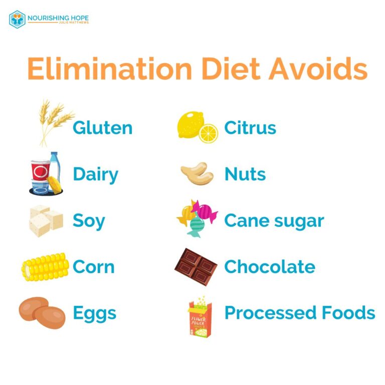
- A review of what actually sustains inflammation: excess free sugars, industrial trans fats, chronic protein deficiency, deficiency of iron, magnesium, B12.
- Assessment of glycaemia. Insulin resistance and neuroinflammation go together, and this is a separate knot worth untying jointly with an endocrinologist.
Biomechanics
Mechanics seems out of place in this list until you look at what exactly feeds glial activation.
It feeds on the flow of nociceptive signals from the periphery. Any constant source of irritation, an overloaded joint, a compressed nerve, a muscle tense for years, keeps the dorsal horn in a state of heightened readiness for years.
Here we recall hypermobility again. A joint with an excessive range of movement needs constant muscular stabilisation, because the ligaments do their job poorly. The muscles work overtime, proprioception is blurred, the position of the body in space is read inaccurately. Hence both chronic fatigue and a tendency to microtrauma. For people with ADHD and other forms of neurodivergence, in whom hypermobility occurs more often, this is one of the main sources of pain.
The second mechanical storyline is immobility. A muscle that does not contract does not merely weaken. It stops releasing myokines. The work of Bente Pedersen showed that a contracting muscle discharges interleukin-6, which in this context behaves as an anti-inflammatory signal and triggers the production of interleukin-10 and of the interleukin-1 receptor antagonist. That is, movement is not a matter of burning calories, it is an endocrine event with an anti-inflammatory effect.
There are data on connective tissue itself as well. In experiments by Helene Langevin’s group, static stretching in rats reduced the volume of the inflammatory focus and accelerated its resolution. Slow stretching may influence not only the length of the muscle but also the local inflammatory process.
And a third point. Diaphragmatic breathing raises vagal tone. We return to the cholinergic anti-inflammatory pathway, but this time from the side of chest biomechanics.
Movement should be regular, dosed and tolerable.
Lifestyle
Sleep, light and rhythm are not healthy habits. They are instruments of direct action on the very same substances that we otherwise try to correct pharmacologically.
Sleep. A meta-analysis by Michael Irwin and co-authors showed a stable association between sleep disturbance and raised inflammatory markers, including interleukin-6 and C-reactive protein. The glymphatic system works separately: according to the group of Maiken Nedergaard (Science, 2013), during sleep the interstitial space of the brain expands and the clearance of metabolic products accelerates sharply. The brain washes itself at night.
Nora Volkow and co-authors showed that sleep deprivation reduces the availability of D2 and D3 dopamine receptors in the striatum. That is, a single night of poor sleep reproduces a dopamine profile similar to the one we discuss in ADHD. A person with chronic pain almost always sleeps badly. After that their attention suffers, they acquire a suspicion of attention deficit, and nobody checks their sleep.
Movement. Besides myokines, regular aerobic exercise produces the phenomenon known as exercise-induced hypoalgesia: a temporary rise in the pain threshold after work. In people with central sensitisation this mechanism often works less well, and one of the goals of rehabilitation is to restore it.
Light and rhythm. Bright morning light into the eyes, ideally within the first hour after waking, synchronises the circadian rhythm and works on evening melatonin better than melatonin itself in a tablet. Melatonin, incidentally, is not only a sleep aid, it has its own anti-inflammatory and antioxidant activity.
Meal timing. Not only composition, but also distribution over time. Eating at night shifts the metabolic rhythms of the liver and adipose tissue, and this is a separate contribution to systemic inflammation.
Alcohol. It increases the permeability of the intestinal barrier and breaks the architecture of sleep, depriving a person of slow-wave phases.
Conclusion
Unlike acute inflammation, chronic inflammation, neuroinflammation included, is not a strong fire but rather an extinguishing that never happened. Not an excess of the attack signal, but a deficit of the all-clear signal.
This understanding completely changes the approach to my recommendations:
- Omega-3 turns from a mere healthy fat into the raw material for the molecules that give the command to stop inflammation.
- Sleep is needed not only for rest, but also for a kind of self-cleaning of the brain.
- Movement is needed not for burning calories, but because a contracting muscle sends the immune system an anti-inflammatory message.
- Gut bacteria are not a fashionable topic, butyrate maintains the intestinal barrier and lowers the level of inflammation.
As for chronic pain, obesity, ADHD and autism sitting side by side in one article, this stops looking like a stretch as soon as it becomes clear that all four conditions have to do with the way microglia treat neurons.
A person with chronic pain and difficulties with attention most probably suffers not from two different problems but from one that has two faces. And treating it in pieces, with four specialists each of whom sees his own quarter, works noticeably worse than treating it whole.