Knee pain. Evidence-based medicine vs common practice
Knee pain. Evidence-based medicine vs common practice
Knee pain is a very common complaint in patients over 40. At the first complaints the usual advice is nonsteroidal anti-inflammatory drugs (Ibufen, Nidometacin, Etopan, Arcoxia), then physiotherapy. Usually this does not help, or helps only a little (I will write separately about why physiotherapy so often turns out to be useless). After treatment fails, an examination is ordered: X-ray, ultrasound and, eventually, MRI. The conclusion after all the tests: “degenerative changes”, “cartilage thinning”, “horizontal tear of the posterior horn of the medial meniscus”.
From this follows the verdict: there it is, the cause of the pain, and it needs fixing. But that verdict is wrong more often than not.
A worn joint is the norm of mature age
Degenerative changes in the knee after thirty-five or forty are not a pathology but an expected age-related finding, like grey hair or wrinkles.
A meta-analysis of 63 studies, almost five thousand people, published in the British Journal of Sports Medicine in 2018, showed that in people with no complaints and no injuries such changes are widespread. Cartilage defects are found in 11% of people under 40 and already in 43% after 40, meniscal tears in 4% under 40 and in 19% after 40.1
So in nearly half of healthy people over forty the cartilage on imaging is not perfect, and it does not stop them living without pain.
A study of 230 knees in ordinary sedentary adults with a median age of 44 (3-tesla MRI, Skeletal Radiology, 2020) took the point to its limit: some abnormality in the knee joints was found in 97% of those examined, meniscal tears in 30%.2
A knee with not a single radiographic finding after the age of forty is a rarity, not a benchmark.
And finally the Framingham study (New England Journal of Medicine, 2008), which every orthopedist cites. Among people aged 50 to 90 meniscal tears were very common, but 61% of those with a torn meniscus had no pain, no aching and no stiffness.3
Changes in meniscal structure, including tears, are most often symptom-free.
The missing link between the imaging picture and the clinical state
A systematic review by Bedson and Croft (BMC Musculoskeletal Disorders, 2008) brought together dozens of studies. Among people with knee pain, radiographic osteoarthritis is found in 15 to 76% of cases, while among those whose osteoarthritis is visible on imaging, pain is present in only 15 to 81%.4 The spread is enormous, the correlation unconvincing.
Images do not reflect the pain.
Both when radiographic changes are present and when they are absent, a large role in the pain is played by so-called central sensitization, a re-tuning of the nervous system’s own pain-processing machinery. A study in Arthritis & Rheumatism (2013) showed that signs of central sensitization are especially pronounced in patients with a high level of pain in the absence of moderate or severe changes on X-ray.5
The pain is entirely real, but its cause is not in worn cartilage and not in degenerative changes of the joint.
Why do knees hurt?
Like any pain syndrome, knee pain has many causes at once. But two of them I would call the main ones.
The first is the state of the muscles. Weakness of the quadriceps is one of the earliest signs, appearing even before the pain. A review in Frontiers in Physiology (2021) states plainly that muscle weakness is characteristic of patients with knee osteoarthritis and predicts loss of function better than pain or joint space narrowing.6
Muscle strength turns out to be a more reliable marker than the state of the joint on X-rays and MRI.
The second cause is chronic subclinical inflammation. Modern rheumatology has effectively rewritten the definition of osteoarthritis. A review in Nature Reviews Rheumatology (2016) puts it this way: osteoarthritis was long considered a degenerative disease of cartilage, but accumulated data show that inflammation plays a key role, and this inflammation is chronic, comparatively low-grade and driven by innate immunity.7 The same belongs here: age-related smouldering inflammation, inflammaging, and inflammation of the synovial membrane. Synovitis is linked both to more pronounced pain and to faster cartilage loss.8
Inflammatory knee pain is not “wear of parts” but an active biological process that can be influenced.
Treating knee pain
Here the evidence is almost unequivocal, and it clearly runs against the usual surgical logic.
The Norwegian OMEX study (BMJ, 2016) enrolled 140 middle-aged patients with a degenerative meniscal tear, and in 96% there were no clear radiographic signs of osteoarthritis.9
Twelve weeks of exercise therapy gave the same result as arthroscopic meniscal resection.
Ten-year follow-up found no difference either. The ESCAPE study (five-year follow-up) confirmed it: arthroscopic meniscal resection has no clinically meaningful advantage over exercise therapy.10
The strictest form of evidence is comparison with a sham operation. The Finnish FIDELITY study (New England Journal of Medicine, 2013) compared a real meniscal resection with an imitation operation, in which incisions are made but the meniscus is not touched, in 146 people aged 35 to 65. The result was the same. The ten-year follow-up of the same study, published in April 2026, went further: surgery gave no advantage over sham surgery, and in the surgery group there was a trend toward worse long-term outcomes, including more symptoms, more pronounced progression of osteoarthritis and more frequent repeat procedures.11
This is not news but a confirmation of earlier studies. Moseley’s work (New England Journal of Medicine, 2002) already showed that in knee osteoarthritis the outcomes after arthroscopic lavage or debridement are no better than after a placebo procedure.12 The Kirkley study (New England Journal of Medicine, 2008) repeated this in another group: adding arthroscopy to physical and drug therapy gave no additional benefit.13
The international OARSI guidelines (2019) place patient education and structured land-based exercise programs at the core of treatment.14
This is not theory. The GLA:D program, grown from these principles, gathered data on 28,370 patients in Denmark, Canada and Australia: people with symptomatic osteoarthritis of the knee and hip show significant improvements in pain intensity, function and quality of life after a course of intensive physiotherapy.15 An important detail: in this program the diagnosis is made clinically, without imaging, and only about 10% of patients with knee osteoarthritis proceed to joint replacement within two years.16
The choice: should you have an MRI for knee pain?
If MRI changes are present in half of healthy peers, and the best results come from strengthening muscles and treating inflammation rather than from surgery, the question arises: what role does imaging itself play. The answer is an uncomfortable one: most often an excessive role.
A register-based study from Norway gives figures every patient should know.
Fewer than 1% of knee MRI results lead to a change in management, and 40 to 95% of such examinations are deemed inappropriate or unnecessary.17
Meanwhile the number of scans keeps growing: in Australia, between 2003 and 2017, the number of knee MRIs increased sevenfold.17
For a typical patient the diagnosis is made without any equipment at all. In people over 45 with activity-related pain and morning stiffness under half an hour, imaging is usually not needed unless there are atypical symptoms.18
When a scan is justified, the X-ray comes first, not the MRI, because a plain radiograph is considered the first-line examination in osteoarthritis.19
The international Choosing Wisely campaign has explicitly placed routine knee MRI for degenerative complaints on the list of procedures that bring no benefit and may cause harm.
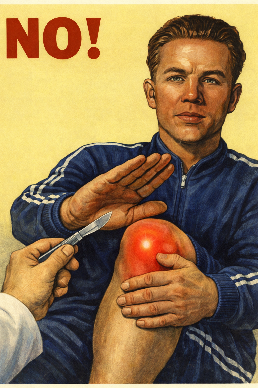
The harm here is not abstract. An MRI will almost certainly find “degeneration” or a “tear” that is present in many symptom-free people of the same age. The patient takes fright, the doctor gains a reason to intervene, then comes arthroscopy, which, as we have seen, works no better than placebo and over the long term may worsen the outcome. A scan does not treat the knee. It sets off a chain of anxiety and unnecessary interventions.
Knee pain is not a verdict on the joint
In a person of mature age, knee pain is in most cases not a signal that the joint is mechanically damaged and needs repair. Degenerative changes are a background, normal for the age. The real causes of pain are different: weakness and imbalance of the muscles, chronic low-grade inflammation and a re-tuning of the pain-perception system. Understanding these causes leads to the right choice of treatment: movement, muscle strengthening, competent physiotherapy and control of inflammation, not a scan and not a scalpel.
Movement, muscle strengthening, competent physiotherapy, control of inflammation.
Routine MRI for degenerative complaints and arthroscopy, no better than placebo.
References
All sources are open via the links; the in-text markers point here.
- Prevalence of knee osteoarthritis features on MRI in asymptomatic uninjured adults: a systematic review and meta-analysis. British Journal of Sports Medicine, 2019.
pmc.ncbi.nlm.nih.gov/articles/PMC6837253 - Prevalence of abnormal findings in 230 knees of asymptomatic adults using 3.0 T MRI. Skeletal Radiology, 2020.
ncbi.nlm.nih.gov/pmc/articles/PMC7237395 - Englund M. et al. Incidental Meniscal Findings on Knee MRI in Middle-Aged and Elderly Persons. New England Journal of Medicine, 2008.
nejm.org/doi/full/10.1056/NEJMoa0800777 - Bedson J., Croft P.R. The discordance between clinical and radiographic knee osteoarthritis. BMC Musculoskeletal Disorders, 2008.
pubmed.ncbi.nlm.nih.gov/18764949 - Finan P.H. et al. Discordance between pain and radiographic severity in knee osteoarthritis. Arthritis & Rheumatism, 2013.
onlinelibrary.wiley.com/doi/abs/10.1002/art.34646 - Benefits and Mechanisms of Exercise Training for Knee Osteoarthritis. Frontiers in Physiology, 2021.
frontiersin.org/journals/physiology/articles/10.3389/fphys.2021.794062/full - Robinson W.H. et al. Low-grade inflammation as a key mediator of the pathogenesis of osteoarthritis. Nature Reviews Rheumatology, 2016.
nature.com/articles/nrrheum.2016.136 - Inflammation in osteoarthritis: our view on its presence and involvement in disease development over the years. Osteoarthritis and Cartilage, 2023.
oarsijournal.com/article/S1063-4584(23)01020-8/fulltext - Kise N.J. et al. Exercise therapy versus arthroscopic partial meniscectomy for degenerative meniscal tear (OMEX). BMJ, 2016.
pmc.ncbi.nlm.nih.gov/articles/PMC4957588 - Physical Therapy vs Arthroscopic Partial Meniscectomy (ESCAPE), five-year follow-up.
ncbi.nlm.nih.gov/pmc/articles/PMC9270699 - Sihvonen R., Kalske R. et al. Arthroscopic Partial Meniscectomy for Degenerative Tear, 10-Year Outcomes (FIDELITY). New England Journal of Medicine, 2026.
medicalresearch.com/nejm-arthroscopic-partial-meniscectomy - Moseley J.B. et al. A Controlled Trial of Arthroscopic Surgery for Osteoarthritis of the Knee. New England Journal of Medicine, 2002.
pubmed.ncbi.nlm.nih.gov/12110735 - Kirkley A. et al. A Randomized Trial of Arthroscopic Surgery for Osteoarthritis of the Knee. New England Journal of Medicine, 2008.
nejm.org/doi/full/10.1056/NEJMoa0708333 - OARSI guidelines for the non-surgical management of knee, hip, and polyarticular osteoarthritis, 2019. Guideline summary.
emedicine.medscape.com/article/330487-guidelines - Outcomes following the GLA:D program, 28,370 patients in Denmark, Canada and Australia. Osteoarthritis and Cartilage, 2020.
oarsijournal.com/article/S1063-4584(20)30118-7/fulltext - GLA:D program outcomes for hip and knee osteoarthritis. Review of GLA:D registry data.
dralisongrimaldi.com/blog/glad-program-outcomes-for-hip-and-knee-oa - Low-value MRI of the knee in Norway: a register-based study.
pmc.ncbi.nlm.nih.gov/articles/PMC10941154 - Use of knee MRI to assess osteoarthritis, room for improvement? ScienceDirect, 2026.
sciencedirect.com/science/article/pii/S1553725026000632 - Unnecessary magnetic resonance imaging of the knee. ScienceDirect.
sciencedirect.com/science/article/pii/S2049080121006865